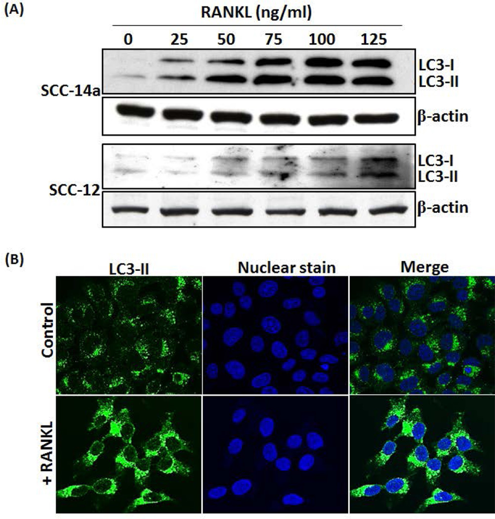
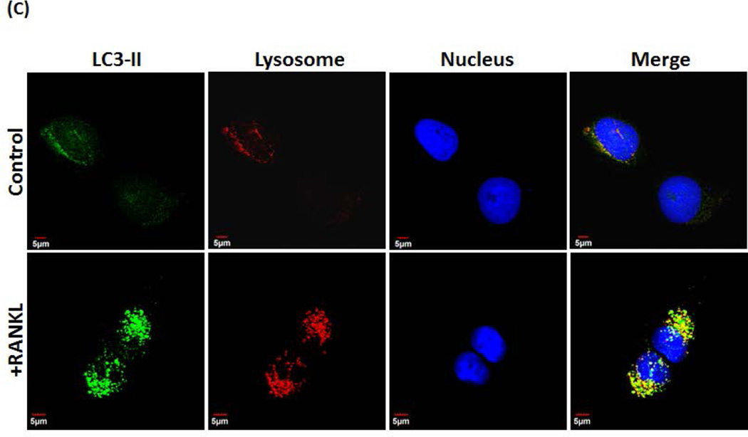
Fig. 4. RANKL increases LC3-I, LC3-II and autophagosome formation in OSCC cells.
(A) SCC-14a and SCC-12 cells were stimulated with different concentrations of RANKL (0–125 ng/ml) for 24 h and total cell lysate were subjected to western blot analysis for LC3 proteins using the anti-LC3 antibody which recognize both LC3-I and LC3-II molecules. β-actin expression served as control. (B) Confocal microscopy analysis of autophagosome formation in OSCC cells. SCC14a cells were stimulated with RANKL (100 ng/ml) for 24 h and immunostained for LC3-II. Nuclear staining was performed by DRAQ5. Merged image demonstrates cytosolic localization of autophagosomes. (C) Co-localization of LC3-II in lysosome. SCC14a cells were stimulated with RANKL and immunostained for LC3-II. Also, label lysosome using LysoTracker® Red DND-99, a red-fluorescent dye. Nuclear staining was performed by DRAQ5. Representative images of cells demonstrate co-localization of lysosome with LC3-II (magnification 60×).


