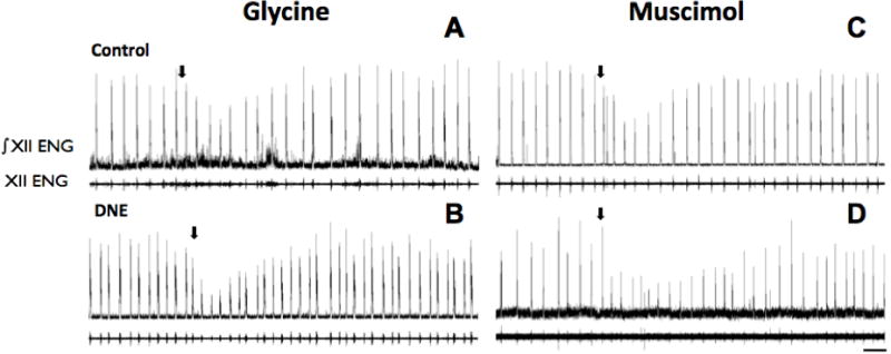
Figure 4. Examples of the nerve burst response to microinjection of muscimol or glycine into the XIIn.

In each panel, the upper trace is the rectified and integrated nerve burst (∫ENG), with the unprocessed burst below. Panels A and C are from control animals, and Panels B and D from DNE animals. Arrows mark the onset of glycine (25 mM for 20 sec; Panels A and B) or muscimol (10 μM for 20 sec; Panels C and D) injection. Note that burst amplitude changed, but there was little, if any influence on burst frequency. Scale bar in lower right panel represents 20 seconds, and applies to all panels in figure.
