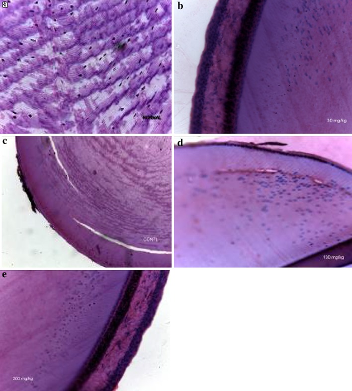
Fig. 6.
Photomicrographs showing normal and selenite-induced cataractous rats treated with various doses of HIE or normal saline (control). a Normal lens showing regular arrangement of equatorial sections of outer lens cortex lens fiber interspersed with nuclei. b Selenite-induced cataractous lens with 30 mg kg−1 HIE treatment showing intact cuboidal epithelium of the lens surface, normal subcapsular epithelial margin, and lens fiber nuclei. c Selenite-induced cataractous lens with 10 ml kg−1 normal saline treatment (control) showing a virtually eroded epithelial margin and distorted lens fiber morphology. d Selenite-induced cataractous lens with 100 mg kg−1 HIE treatment indicating a partially eroded epithelium and moderate mild changes in lens fiber morphology. e Selenite-induced cataractous lens with 300 mg kg−1 HIE treatment showing preserved cuboidal epithelium of the lens surface and normal subcapsular epithelial margin. HIE whole plant extract of Heliotropium indicum

