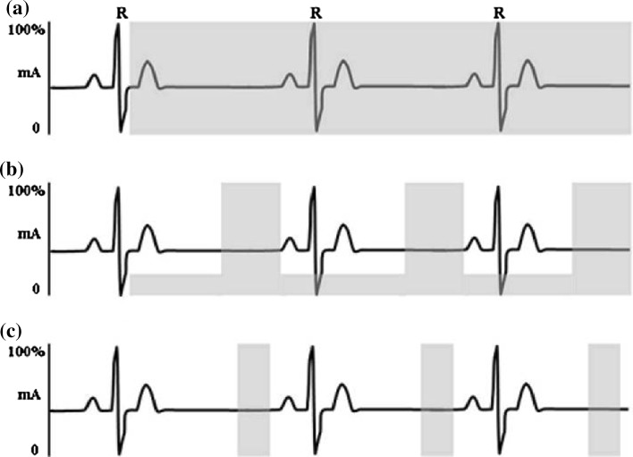
Fig. 4.
Diagram illustrating image acquisition (and thus radiation exposure) during the different types of gating. a Full-dose retrospective gating, with a constant high level of radiation. b Electrocardiogram tube dose modulation, with a constant low level of radiation, which is increased during mid-diastole when the main part of the image is acquired. c Prospective imaging during which the image acquisition (and thus radiation exposure) occurs only at pre-set intervals, again generally during mid-diastole. Reproduced from Weustink and de Feyter [89]. This article was published under the Creative Commons Attribution Non-Commercial License

