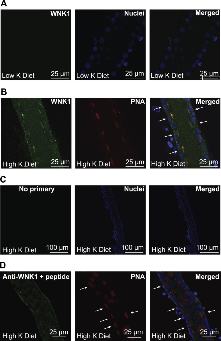
Fig. 6.
High-K+ (HK) diet increases L-WNK1 expression in rabbit cortical collecting ducts (CCDs). Single CCDs isolated from New Zealand White (NZW) rabbits fed a HK or a low-K+ (LK) diet for 10 days were microperfused and fixed for immunofluorescence microscopy. Immunolabeling was performed as indicated in materials and methods. A: L-WNK1 expression in CCDs from LK-fed animals. Immunostaining for L-WNK1 was difficult to visualize in CCDs from rabbits fed a LK diet. B: expression of L-WNK1 in rabbit CCD intercalated cells after 10 days on a HK diet. Intercalated cells (arrows) were identified by their apical labeling with rhodamine-conjugated peanut agglutinin (PNA). C: negative control incubated with Alexa488-conjugated goat anti-rabbit IgG antibody but not with primary antibody. D: antigen competition. Only modest basal membrane labeling was seen when the anti-L-WNK1 antibody was preincubated with a WNK1 peptide fragment (arrows indicate PNA+ cells).

