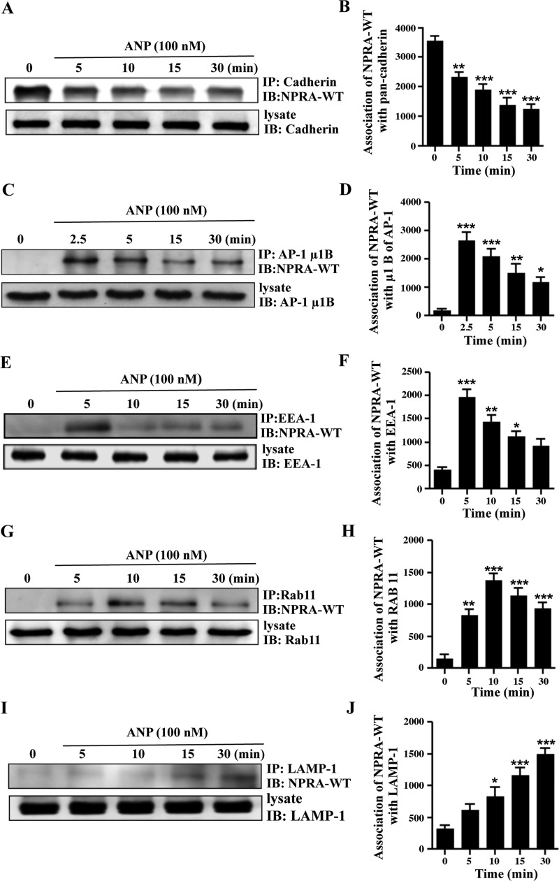
Fig. 9.
Coimmunoprecipitation of eGFP-NPRA-WT with pan-cadherin, μ1B, EEA-1, Rab 11, and LAMP-1 in MMCs. To determine the association of NPRA with pan-cadherin, μ1B, EEA-1, Rab 11, and LAMP-1, cells were stimulated with 100 nM ANP for different time points. Anti-eGFP antibody was utilized for immunoblotting (IB) of eGFP-NPRA fusion protein. A: immunoblot of NPRA after immunoprecipitation (IP) of pan-cadherin showed a decreased association with increasing time points. B: densitometric Western blot quantification of NPRA association with pan-cadherin relative to untreated cells. C: coimmunoprecipitation of NPRA with μ1B showed maximum association at 2.5 min, and after that it gradually decreased. D: densitometric Western blot quantification of NPRA with μ1B relative to untreated cells. E: coimmunoprecipitation of NPRA with EEA-1 showed maximum association at 5 min, and after that it gradually decreased. F: densitometric Western blot quantification of NPRA with EEA-1 relative to untreated cells. G: strong association of receptor and recycling endosomes was observed at 10 min, and after that it gradually decreased. H: densitometric Western blot quantification of NPRA with Rab 11 relative to untreated cells. I: association of NPRA with lysosomes gradually increased after 5 min, and it was maximum at 30 min. J: densitometric Western blot quantification of NPRA with LAMP-1 relative to untreated cells. Values are means ± SE of 4 independent experiments. *P < 0.05, **P < 0.01, ***P < 0.001 relative to untreated cells.

