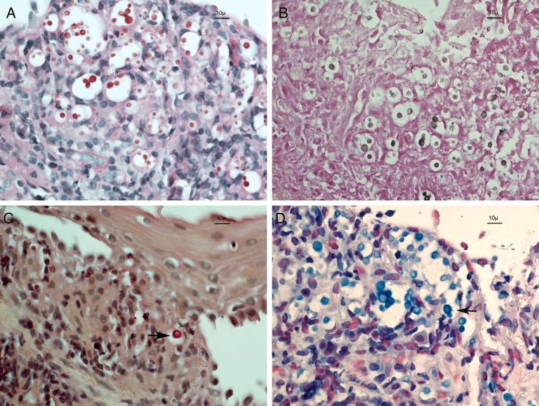
Figure 2.
Four different staining methods of the vocal cord biopsy defined that the infecting yeast was a Cryptococcus neoformans. Pink periodic acid-Schiff-positive yeast (A) take a brown color with the Fontana-Mason staining (B), indicating the presence of melanin. Polysaccharide capsules were stained in pink by the mucicarmine method (arrow, C) and light blue by the alcian blue method (arrow, D). Original magnification, 600×.

