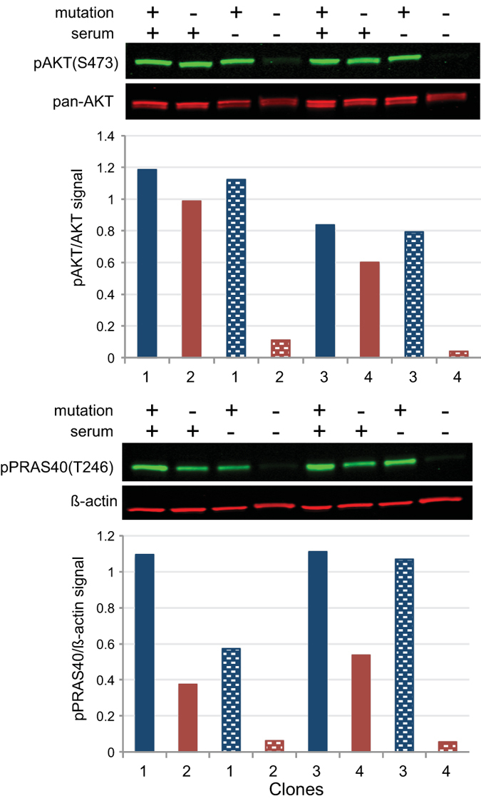
Figure 1. AKT remained active in mutation-positive cells in the absence of growth factors.

Single cell clones isolated from fibroblasts cultured from an epidermal nevus located on the dorsum of the right hand of patient PS134 were grown with and without serum (indicated above gel images) followed by lysis and western analyses. Clones 1 and 3 (blue bars) were positive for the AKT1 E17K mutation; clones 2 and 4 (red bars) were negative. Histograms show ratios of the infrared signals from each antibody pair as described in the methods. Shaded bars represent ratios in cells grown in serum-free medium. Levels of pAKT and pPRAS40 were elevated in the absence of serum in mutation-positive cells compared to mutation-negative cells.
