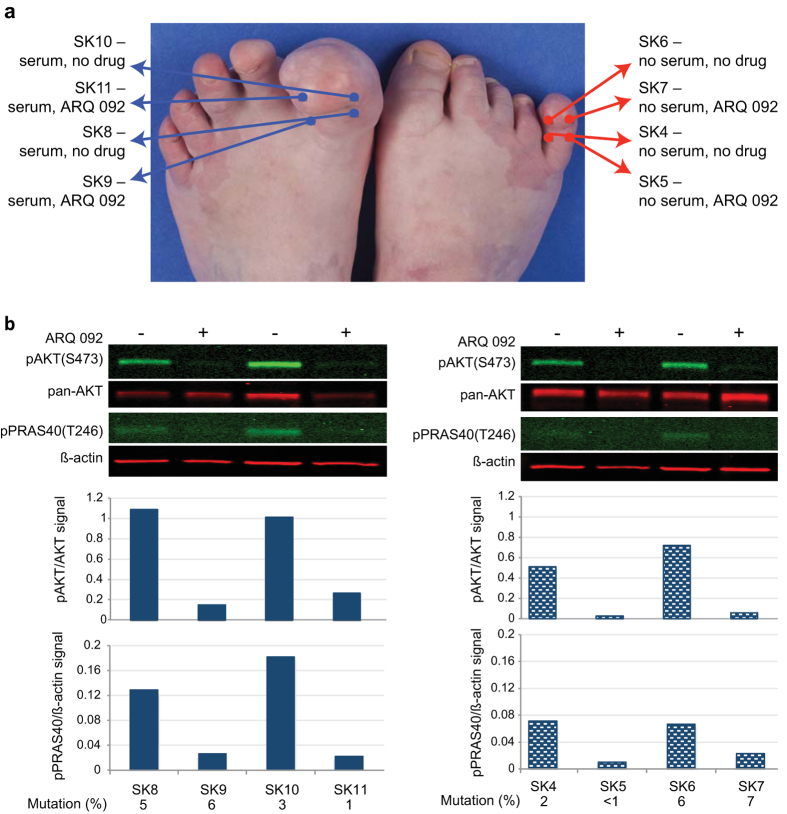
Figure 4. ARQ 092 decreased phosphorylation of AKT and PRAS40 in tissues from a patient with Proteus syndrome.
(a) The location and incubation conditions of punch biopsies taken from two affected toes from patient PS95 are indicated on the photo. Proteins and DNA were extracted as described in Methods. (b) Infrared images of western hybridizations using the indicated antibodies. Histograms below each lane represent the ratio of indicated antibody pair in each lysate. Symbols above images indicate the presence or absence of ARQ 092. AKT1 E17K mutation levels for each biopsy are shown below the graphs.

