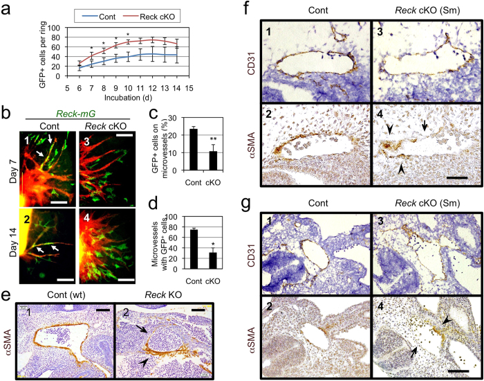
Figure 4. Effects of Reck on vessel-association of Reck-mG cells in ARA and mural cells in vivo.
(a) Time course for the number of Reck-mG cells in ARA. Aortic rings from the 5-weeks-old control (n = 10) or Reck cKO (n = 7) mice were subjected to ARA and all Reck-mG cells surrounding each ring (irrespective or their association with microvessels) were counted from day 6 to 14. (b) Distribution and morphology of Reck-mG cells (green) at day 7 and 14 in ARA. In control cultures, Reck-mG cells are tightly associated with microvessel stalks (arrows in panel 1 and 2) but this is impaired in Reck cKO (panel 3 and 4). (c) Frequency of Reck-mG cells tightly associated with microvessels at day 8. n = 3. (d) Frequency of microvessels with tightly associated Reck-mG cells at day 8. n = 3. (e–g) Effect of Reck-deficiency on the distribution of mural cells in vivo. (e) Sagittal section of wild type (Cont) or Reck−/− (Reck KO) mouse embryos at E10.5 immunostained for αSMA (brown). Micrographs focusing on large aorta are shown. (f,g) Sagittal sections of ReckE1fx/∆ mice (control; panels 1, 2) and Reck cKO (Sm) mice (panels 3, 4) at E10.5. Adjacent sections were immunostained for CD31 (with Hematoxylin counter-stain; panels 1, 3) and for αSMA (panels 2, 4), respectively. Micrographs focusing on large aortas in the middle (f) and caudal (g) part of the body are shown. In (e–g), arrowhead indicates an area of vessel wall abundant in mural cells, and arrow highlights an area where the mural cell layer is very thin or absent. Scale bar: (b) 200 μm (panel 2) and 50 μm (other panels), (e) 100 μm, (f) 50 μm, (g) 100 μm.

