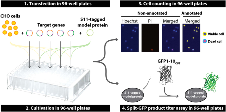
Figure 1. Schematic illustration of the screening platform.
(1) CHO cells are co-transfected with plasmids encoding target genes and plasmids encoding S11(Split-GFP)-tagged model protein. (2) Transfected cells are cultivated in a 96-half-deepwell microplate capped with a low-evaporation Duetz sandwich cover (cover not shown). (3) Viable cell density is determined using image fluorescence cytometry on Hoechst and propidium iodide (PI) stained cells. (4) Upon addition of GFP1–10OPT to supernatants, relative product titers of supernatants are determined by split-GFP complementation.

