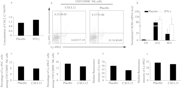
Figure 5. CXCL12 did not modulate the percentages of Ly-49+ NK cells in vitro.
(a) Syngeneically mated BALB/c females were injected with placebo or IFN-γ intraperitoneally on GD6 and sacrificed on GD8. CXCL12 concentration in the serum was determined by ELISA. Data show the mean ± SEM of four independent experiments and were obtained from four mice per group. (b–f) Splenic leucocytes were cultured with CXCL12 at a dose of 500 ng/ml for 24 h. (b) Representative flow cytometric analysis of Ly-49A and Ly-49G2 expression on gated CD3−CD49b+ NK cells; the numbers in the dot plots indicate the percentages of the Ly-49+ NK cells (gated on CD3−CD49b+ NK)/MFI values of Ly-49 receptors expression. Data summary of the percentages of Ly-49+ NK cells (c,d) and MFI values of Ly-49 receptor expression (e,f). Data show the mean ± SEM of three independent experiments. (g) Splenic leucocytes were cultured with IFN-γ at a dose of 250 U/ml and at the indicated times, splenic leucocytes were harvested to determine CXCR4 expression by quantitative PCR. Data show the mean ± SEM of four independent experiments.

