
Figure 1. Atrial Tachycardias.
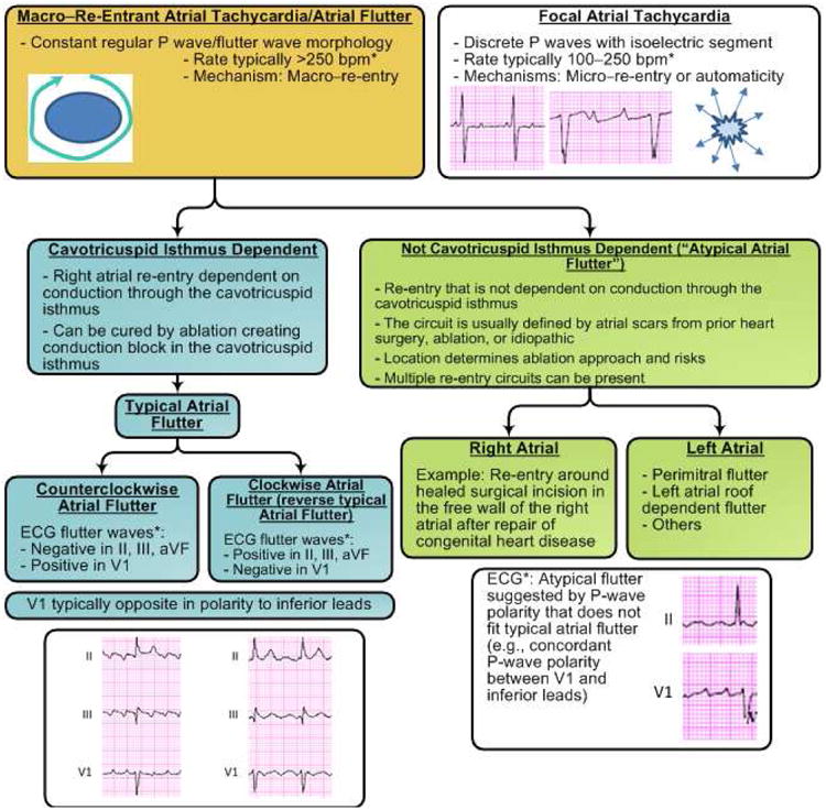
Diagram summarizing types of atrial tachycardias often encountered in patients with a history of AF, including those seen after catheter or surgical ablation procedures. P-wave morphologies are shown for common types of atrial flutter; however, the P-wave morphology is not always a reliable guide to the re-entry circuit location or to the distinction between common atrial flutter and other macro–re-entrant atrial tachycardias.
*Exceptions to P-wave morphology and rate are common in scarred atria.
AF indicates atrial fibrillation and ECG, electrocardiogram (72, 80).