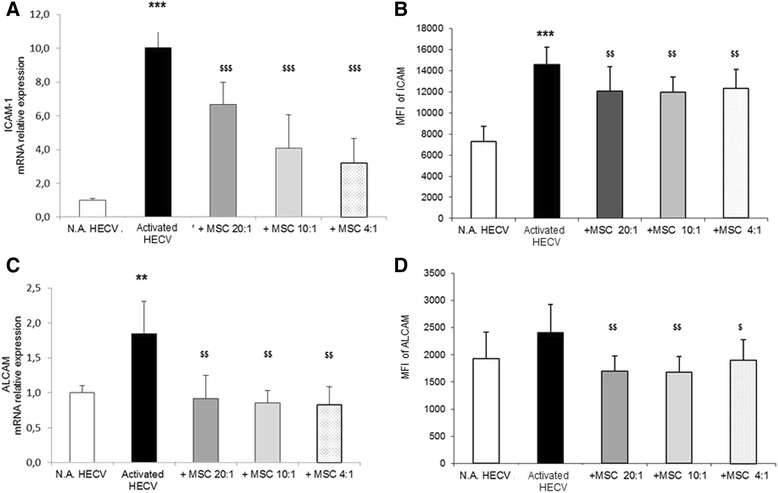
Fig. 4.

Effects of MSC on expression of HECV adhesion molecules. HECV stimulated using IFNγ were cultured for 24 hours in the absence or in the presence of MSC at the HECV/MSC ratios of 20:1, 10:1, and 4:1 in TW. The relative mRNA expression of ICAM-1 a and ALCAM c were evaluated by qPCR. Data (mean ± SD) are reported as fold induction with respect to controls. In the same experimental conditions, the mean fluorescence intensity (MFI) of plasma membrane expression of ICAM-1 b and ALCAM d was evaluated by flow cytometry following staining with specific mAb. Results of six independent experiments are expressed as mean percentage ± SD of recorded MFI. Activated vs. N.A. cells, *p ≤0.01, **p ≤0.001, ***p ≤0.0001; MSC-treated vs. MSC-untreated cells, $p ≤0.01, $$p ≤0.001, $$$p ≤0.0001. ALCAM activated leucocyte cell adhesion molecule, HECV human endothelial cells, ICAM intercellular adhesion molecule, MSC mesenchymal stem cells, N.A. PBMC in resting conditions
