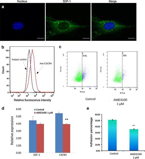
Fig. 4.

a 3D confocal images of SDF-1 staining on M210B4 stromal cells in X–Y plane. The scale bar is 10 µm. b Surface expression of CXCR4 on Molm13 cells. Molm13 cells were incubated with anti-CXCR4 and labeled with an FITC conjugate. Controls received equivalent concentrations of isotype-matched IgG. Washed cells were analyzed by flow cytometry, in which accumulated events were gated against the isotype control. c Flow cytometer tested the expression of CXC4 in molm13 cells. The CXCR4 expression cells decreased after 1 μM AMD3100 treated. d Expression of sdf-1has no significant difference after 1 μM AMD3100 treated in M210B4 cells. Cxcr4 expression significantly decreased after AMD3100 treated (p < 0.05). e Characterization of adhesion of Molm13 cells on M210B4 cells with and without drug treatment. Molm13 cells were treated with 1 µM AMD3100 for 2 h before experiments. Experiments were repeated for eight times. *P < 0.05. ***P < 0.001
