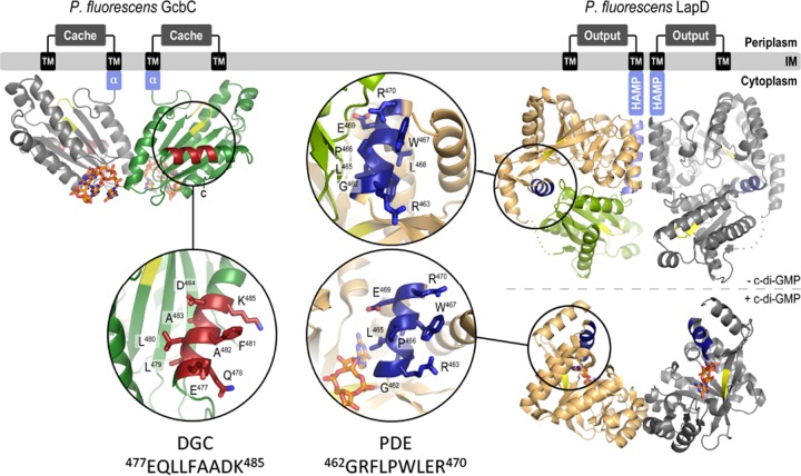
FIG 6 .
Model for GcbC-LapD interactions. (Left) GcbC bound to c-di-GMP in inactive dimer. The inset shows the α5GGDEF helix (residues 477 to 485) in red. (Right) LapD in its unbound state (above) and c-di-GMP-bound, active state (below). Insets show the α2EAL helix of LapD in dark blue. The helix is exposed in the c-di-GMP-bound state of LapD.

