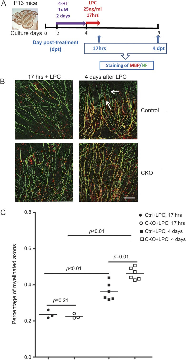
Figure 4. CXCR2 deficiency on oligodendrocyte lineage cells causes rapid and more complete remyelination in an in vitro model of demyelination/remyelination using LPC-treated cerebellar slice cultures.

(A) Schematic diagram for slice culture on control and cKO mice. (B) Cerebellar slice from lysophosphatidylcholine (LPC) treated (17 hours) and 4 days after LPC treated from control and cKO mice were collected and dual labeled with myelin basic protein (MBP) (red) and neurofilament (green), with myelinated fibers appearing yellow in merged images. At 17 hours, all slices are demyelinated, showing abundant green unmyelinated nerve fibers and scant yellow myelinated fibers. Four days after removing LPC, better remyelination is shown in slices from cKO mice compared to control mice. (C) The customized Fiji software used to quantify the colocalization of myelin sheath on axons as percentage of myelinated axons is described in detail in the Methods. Scale bar, 25 μm. Arrow: Unmyelinated axons. These data represent 5 independent experiments. Each experiment included 3–4 mice each group.
