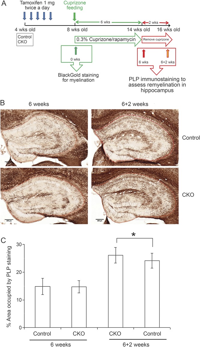
Figure 5. CXCR2 deficiency on oligodendrocyte lineage cells accelerates remyelination in a cuprizone/rapamycin-induced demyelination model.
(A) Schematic diagram for cuprizone feeding on control and Cxcr2-cKO mice. (B) Control and Cxcr2-cKO mice 4 weeks after tamoxifen injection were fed cuprizone diet and received daily IP rapamycin injections for 6 weeks. Tissues were collected after cuprizone feeding for 6 weeks or after 2 weeks recovery from 6 weeks cuprizone. PLP antibody staining for myelin shows equivalent demyelination severity of the hippocampi in tamoxifen-treated control and Cxcr2-cKO mice at 6 weeks of cuprizone/rapamycin treatment. Tamoxifen-treated Cxcr2-cKO mice show significantly accelerated remyelination at 2 weeks compared to tamoxifen-treated control mice. Scale bars: 200 μm. (C) Percent of PLP occupied area in the hippocampal area after cuprizone treatment shows no significant differences between these 2 groups at 6 weeks feeding. Percent of PLP occupied area in the hippocampal area shows significant differences between tamoxifen-treated control and Cxcr2-cKO mice remyelination, 2 weeks post recovery from 6 weeks demyelination. *p < 0.05. n = 8 per group for 6 weeks demyelination; n = 15 per group for 2 weeks remyelination.

