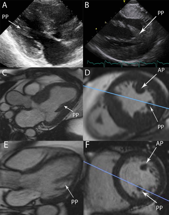
Figure 5.

(A) shows a typical TTE PLAX echo image, the papillary muscle in view is the posterior muscle. (B) shows a typical ICE long-axis view of the left ventricle, with both heads of the posterior muscle visible. (C) shows a three-chamber CMR still with a papillary muscle labelled. Relational short axis CMR images show this to be the posterior muscle (PP), the blue line represents the plane displayed in the three chamber view in (C). The anterior muscle (AP) is out of shot and would be very difficult to see in the same plane as the LVOT. The same perspectives are displayed in the non-HCM heart in (E) and (F).

 This work is licensed under a