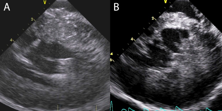
Figure 7.
Both (A) and (B) show ICE images in systole with SAM-septal contact. In each example, the AMVL is folded back and has a bend of nearly 90. In (B), an area of iatrogenic infarct in the mid-septum from previous ASA is seen. The contact area for the AMVL is further basal, there is viable, contracting myocardium in this area.

 This work is licensed under a