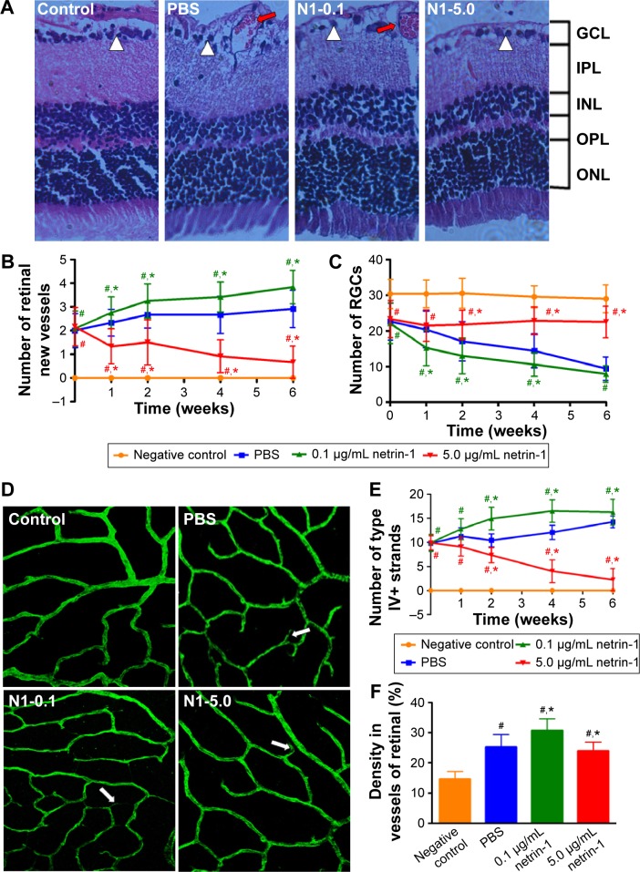
Figure 7.
Retinal structure and vascular changes in the innermost vascular plexus in the four groups.
Notes: (A) The picture of HE staining of whole retinas in the four groups at 6 weeks after intravitreal injection (×100). HE staining of retinal tissues demonstrated that there were no new vessels and sufficient RGCs in the normal rats. After 6 weeks of intravitreal injection with PBS and 0.1 μg/mL netrin-1, the retinal tissues of the diabetic rats contained more new vessels and fewer RGCs. The number of new retinal vessels in the rats injected with 5 μg/mL netrin-1 led to fewer new retinal vessels and more RGCs at 6 weeks after treatment. The large white triangles highlight RGC cells and the red arrows show the new vessels in the retinas. The number of neovascularizations (B) and RGCs (C) were counted and observed. Data are presented as the percentage of nontreated and shown as mean ± SD; n=5, #P<0.05 vs control; *P<0.05 vs PBS. Vascular changes in the innermost vascular plexus were observed by confocal microscopy in the four groups. Six weeks after intravitreal injection, the rats were sacrificed and the retinas were stained with collagen IV (D). Collagen IV staining in the four groups at 6 weeks after injection showed a significant difference between the PBS and netrin-1 (0.1 μg/mL or 5 μg/mL) treated groups. The large arrows showed the collagen IV new strands. Quantification for the number of type IV strands (E) at 6 weeks after intravitreal injection also showed the differences between these four groups. Data are presented as the percentage of non-treated rats and shown as mean ± SD. n=5, #P<0.05 vs CTR; *P<0.05 vs PBS. Quantification for the density of collagen IV (F) at 6 weeks after intravitreal injection also showed the differences between these four groups. Data are presented as the percentage of nontreated rats and shown as mean ± SD; n=5, #P<0.05 vs control; *P<0.05 vs PBS.
Abbreviations: SD, standard deviation; HE, hematoxylin-eosin; PBS, phosphate-buffered saline; RGCs, retinal ganglion cells; GCL, anglion cell layer; IPL, inner plexiform layer; INL, inner nuclear layer; OPL, outer plexiform layer; ONL, outer nuclear layer; CTR, control; PBS, phosphate-buffered saline.

