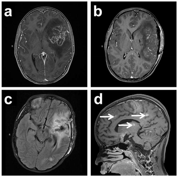
Fig. 1.
Magnetic resonance imaging (MRI) of the brain during the patient’s clinical course. (a). Initial MRI on admission Day 1 revealed a nonspecific multifocal lesion involving the left frontal temporal lobe with surrounding edema and midline shift. (b). MRI on Day 8 revealed postoperative changes with diminished mass effect and midline shift. (c). MRI on Day 20 (flair axial image) revealed a new lesion in left temporo-occipital white matter (arrow) and further reduction in edema with resolution of midline shift. (d). MRI on Day 26 (T1 sagittal image) revealed numerous new foci throughout the brain (arrows)

