Fig. 3.
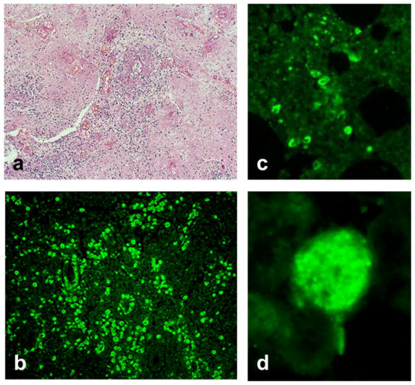
Hematoxylin and eosin (H&E) or indirect immunofluorescence (IFA) staining of tissue sections. (a) A section of brain stained with H&E—Balamuthia amebae are seen interspersed within the tissue at magnification X 100. (b) A similar section of brain reacted with anti-Balamuthia mandrillaris serum in the IFA test at magnification X 100. (c) IFA reactivity of Balamuthia amebae in a section of lung at magnification X 100 indicating dissemination of amebae into the lung. (d) A high power (magnification X 1,000) view of an ameba in the lung by IFA staining

