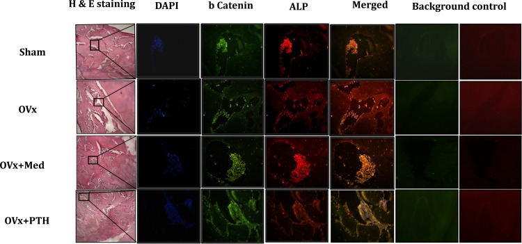
Fig 5. Hematoxylin and eosin staining of newly regenerated bone at the injury site and immunoflourescence analysis of the co localization of β-catenin and ALP in femoral bone sections at the injury site.
Immunofluorescence staining of β-catenin (green) and ALP (red) was performed. Co localization of two molecules is demonstrated in merged yellow image. Cells were counterstained with DAPI (blue). Auto fluorescence images were taken in red and green channel for each panel for normalizing the background.

