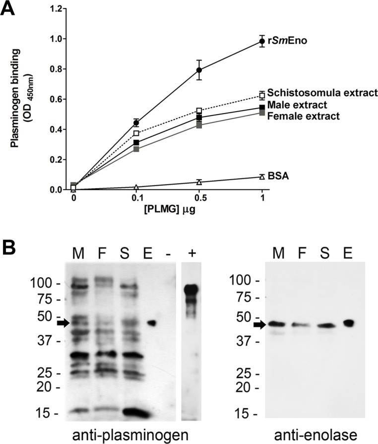
Fig 8. Plasminogen (PLMG) interaction with rSmEno and with schistosome lysates (A) by ELISA or (B) by western blotting.

(A) ELISA plates were coated with rSmEno (0.5 μg/well) or the indicated parasite extracts (1.0 μg/well each) and wells were incubated with increasing concentrations of PLMG (0–1 μg) in triplicate. As a negative control, some wells were coated with BSA (0.5 μg/well). Anti-PLMG antibody was used to detect PLMG following standard ELISA conditions. The lines represent the mean absorbance values at OD 450 nm (± SD). (B) Detection by western blot of schistosome PLMG-binding proteins (left panel). Lanes contain extracts from males (M), females (F) and schistosomula (S), as well as pure rSmEno (E), BSA (“-”, negative control) and commercially-obtained PLMG (“+”, a control for anti-PLMG antibody binding). Multiple bands in the schistosome extracts bind PLMG. The arrow indicates the position of migration of SmEno, here revealed to be a PLMG-binder. No binding to the negative control protein (BSA) is seen. The membrane was stripped and re-probed with anti-ENO1 antibody (right panel). A single, prominent ~47-kDa SmEno band is detected (arrow) in the extracts of males (M), females (F) and schistosomula (S) and in the case of purified rSmEno (E). Images are representative of three replicate experiments.
