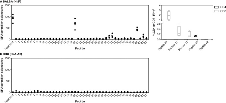
Fig 2. Immunodominant responses to PfLSA1.
(A) BALB/c or (B) HLA-A2 tg mice (n = 5) were vaccinated i.m. with 1x10⁸ ifu ChAd63-PfLSA1 followed eight weeks later by 1x10⁷ pfu MVA-PfLSA1. Two weeks post-MVA boost, mice were sacrificed and splenocytes isolated to perform an ex vivo IFNγ ELISpot. Splenocytes were stimulated with either an overlapping peptide pool to PfLSA1 or individual peptides (20aa each, overlapping by ten). Both median and individual data points are shown. For (A) BALB/c, CD4+ and CD8+ epitopes were also determined (right panel). BALB/c mice (n = 4) were vaccinated with 1x10⁸ ifu ChAd63-PfLSA1 and two weeks later sacrificed and splenocytes isolated. Cells were incubated for six hours with the relevant peptide prior to ICS staining. Box plots show the percentage IFNγ+ of CD4+ or CD8+ cells, with whiskers representing the maximum and minimum.

