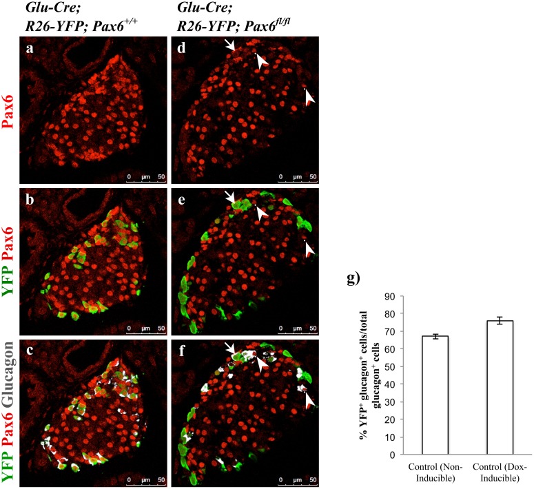
Fig 16. Alpha-cell-specific ablation of Pax6.
Double immunofluorescence staining of pancreatic cryosections from 1 month old mice. In control islets, all the YFP+ cells express Pax6 and glucagon (a-c). In alpha-cell-specific Pax6 KO islets, most of the YFP+ cells are negative for Pax6 and have lost the expression of glucagon as well (d-f). YFP- glucagon+ cells in the KO islets express Pax6 (arrowheads d-f). Rarely YFP+ glucagon+ Pax6- cells are also found in the KO islets (arrows d-f). Quantification of YFP+ glucagon+ cells in relation to total glucagon+ cells in 1 month old non-inducible alpha-cell-specific control (Glu-Cre;R26-YFP;Pax6 +/+) mice, and in Dox-inducible alpha-cell-specific control (Glu-rtTA;TetO-Cre;R26-YFP;Pax6 +/+) mice at 4 months of age following 6 weeks of Doxycycline treatment (g) (n = 3). Nearly 68% and 75% of the glucagon+ cells are labeled with YFP in non-inducible and Dox-inducible alpha-cell-specific control mice, respectively. Error bars represent SEM.

