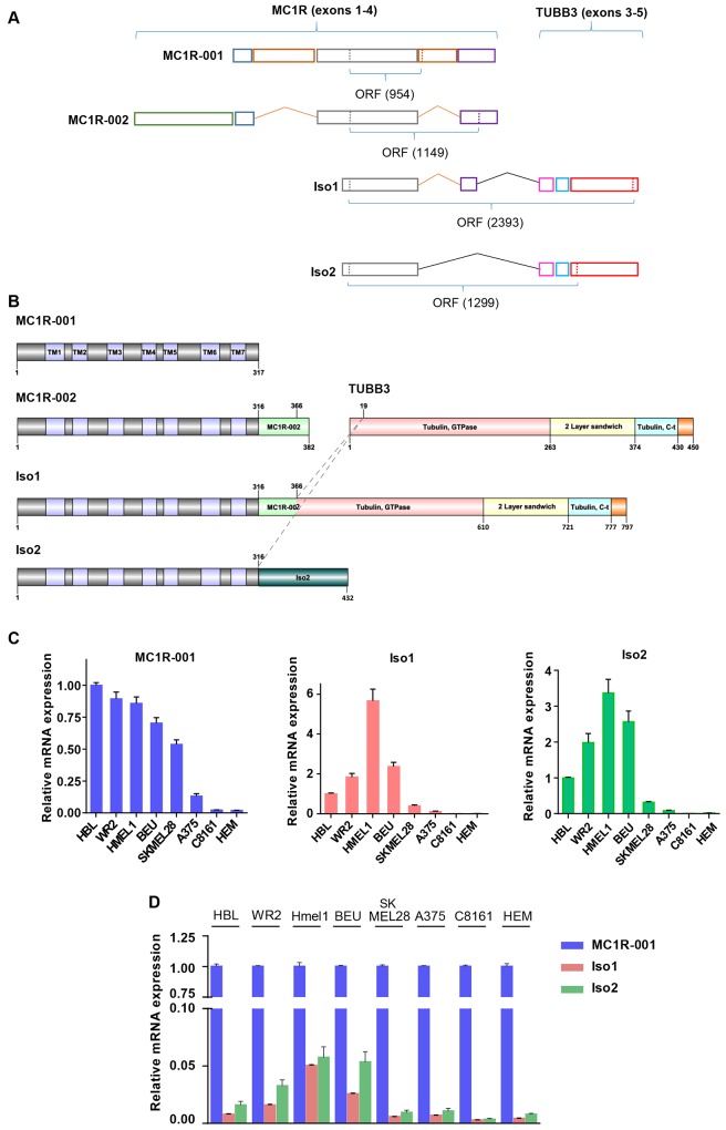
Fig 1. MC1R transcripts and intergenic splice isoforms of MC1R and TUBB3.
(A) Schematic panel showing the exon organization of MC1R splice variants (MC1R-001 and MC1R-002) and MC1R-TUBB3 chimeric transcripts, Iso1 and Iso2. Exons of all MC1R derived transcripts are represented in colored boxes and the number of nucleotides in the ORF is shown below. (B) Diagram representing the structural domains of MC1R-001, MC1R-002, β-tubulin III (TUBB3) and chimeric proteins Iso1 and Iso2. Structural and functional domains are depicted in colored boxes and the number of key residues in the proteins is shown. TM indicates transmembrane regions of MC1R. Dashed lines indicate residues of MC1R and TUBB3 linked in fused proteins Iso1 and Iso2. (C) MC1R-001, Iso1 and Iso2 expression in human melanoma cell lines. Data are shown as relative expression of each isoform (as indicated in each bar graph) as compared with the levels of the isoform in HBL cells. (D) Expression of Iso1 and Iso2 mRNA as a function of the levels of the canonical MC1R-001 transcript in a panel of human melanoma cell lines. Data are represented as mRNA expression of the two intergenic splicing forms relative to MC1R-001 in each cell line.

