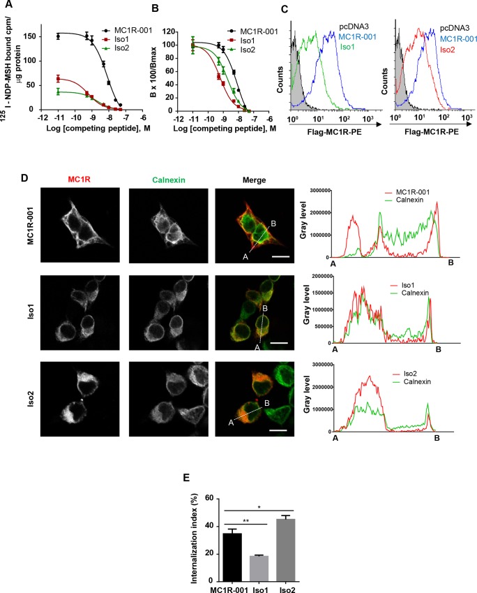
Fig 3. Radioligand binding and intracellular trafficking properties of MC1R-TUBB3 isoforms.
(A-B) Competition binding assay of HEK293T cells transfected with MC1R-001, Iso1 and Iso2. Cells were incubated with 125I-labelled NDP-MSH (5x10-11 M) and increasing concentrations of non-labelled competing NDP-MSH, from 10−12 to 10−7 M, extensively washed and counted for radioactivity. Non-specific binding was determined with non-transfected cells or with transfected cells incubated with the radioactive tracer in the presence of excess (10−6 M) non-labelled peptide, with the same results. Values are represented as specifically bound [125I]-NDP-MSH (A) and as percentage of residual binding (B) at the different ligand concentrations (n = 3, data are given as mean ±SEM). (C) Flow cytometric analysis of HEK293T cells expressing MC1R-001 and MC1R-TUBB3 chimeric isoforms. Non-permeabilized cells expressing the indicated proteins were incubated with an anti-Flag antibody labelled with phycoerythrin. Since the Flag epitope was fused in-frame to the extracellular N-terminus of the MC1R sequence, only cells expressing the constructs of the plasma membrane should be detected. Histograms represent cell number (counts) as a function of Flag surface staining, on a logarithmic scale. The gray filled curve refers to cells transfected with an empty pcDNA3 (n = 3, representative histograms are shown). (D) Left panel: Representative confocal images of MC1R-001 or the chimeric isoforms (red) and calnexin (green) immunostaining in HEK293T cells transiently transfected with Flag-labelled MC1R-001 and MC1R-TUBB3 constructs. Scale bar, 10 μm. Representative line scan (right panel) from multiple experimental repeats across the cell (location indicated in merged image) shows co-localization of MC1R-TUBB3 transcripts and calnexin. Line scan, 19 μm for MC1R-001, 17 μm for Iso1 and 18 μm for Iso2. (E) Radioligand internalization assay performed on HEK293T cells expressing MC1R-001, Iso1 or Iso2 incubated with 125I-labelled NDP-MSH. The radioactive tracer was isotopically diluted to achieve a final concentration of 5x10-11 M and 5x10⁴ counts/well. Externally bound agonist was separated by an acid wash procedure. Both the externally bound ligand present in the acid washes and the internalized ligand associated with the cell pellets were counted. The internalization index represents the percentage of ligand internalized referred to total radioligand bound (n = 3, error bars are ±SEM, two-sided one-way ANOVA was used to generate p values, *p<0.05, **p<0.01).

