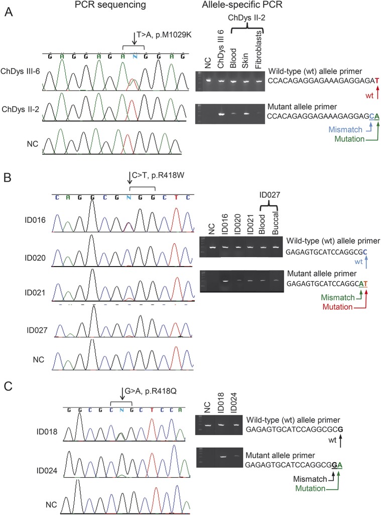
Figure 3. Allele-specific amplification documents mosaicism of ADCY5 mutations in representative cases.
Wild-type (wt) alleles (top panels) were amplified using wt-primer and mutant alleles (lower panels) amplified using primers containing the mutant nucleotide in the 3′ position and one mismatched nucleotide in the preceding position. (A) Sequence chromatograms for 2 members of the ChDys family. The mutant nucleotide A is detected in patient III-6 but not in her affected mother II-2. Allele-specific PCR in II-2 amplified the mutant allele from blood, skin, and fibroblast cells. The analysis is not quantitative, but the very faint mutant band in fibroblasts suggests a selective advantage for the wt allele during culture. (B) On the sequence chromatograms, in contrast to C and T peaks of equal intensity in ID016, much smaller signals are present for the mutant T allele in ID020, ID021, and ID027. Presence of the mutant allele was confirmed for all by allele-specific amplification. (C) Mosaicism for the mutant A nucleotide is shown for ID024, in contrast to the peaks of equal height for the wt and mutant alleles in ID018. In all panels, for the mosaics, the faint gel signals are consistent with the small mutant allele peaks in the chromatograms. Absence of a mutant allele in a normal control (NC) demonstrates the specificity of the allele-specific amplification. ChDys = chorea-dystonia.

