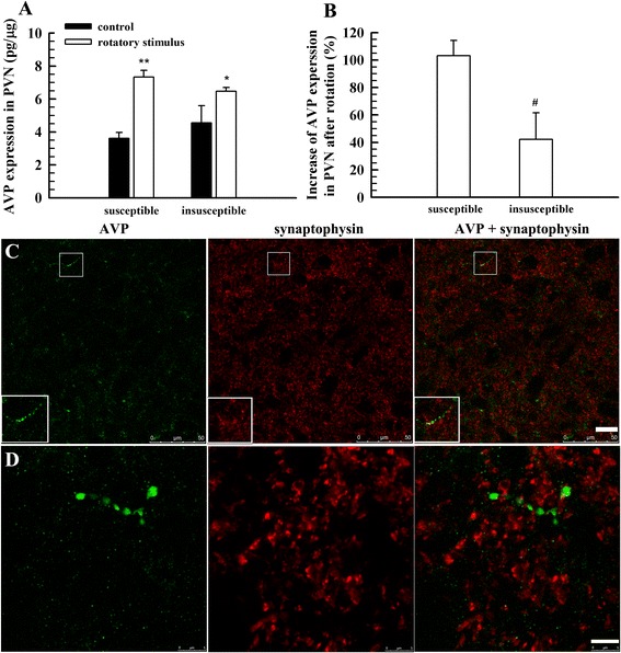
Fig. 5.

The expression of AVP in the PVN and the distribution of AVP-positive fibres in the VN. a, AVP expression in the PVN of susceptible and insusceptible groups with and without rotatory stimulus (n = 6); b, net increase of AVP expression in the PVN after rotatory stimulus (n = 6); c, labelling of AVP and synaptophysin; d, another section labelling AVP and synaptophysin and further magnification to reveal AVP fibre terminals that express synaptophysin. Four rats, two for each gender, were used in (c) and (d). * P < 0.05, ** P < 0.01, vs. control. Scale bar, 20 μm for (c), 5 μm for (d)
