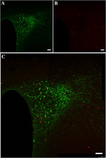
Fig. 6.

Retrograde tracking of AVP fibres from the PVN to the VN. Four rats, 2 for each gender, were used. Serial sections covering the PVN were taken from each animal for observation. a, AVP-positive cells in the PVN; b, fluoro-ruby-labelled cells through retrograde tracking from the VN; c, merge of (a) and (b) and further magnification. Scale bar, 75 μm
