
Fig. 1.
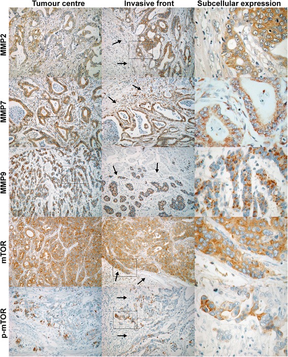
Immunohistochemical staining at the tumor center and the invasive front. Exemplary staining of MMP2, MMP7, MMP9, as well as mTOR and p-mTOR in the tumor center, at the invasive front (100×), and subcellular expression with larger magnification (400×). Arrows are marking the marginal zone of the tumor at the invasive front. Squares are marking the magnified area for each panel