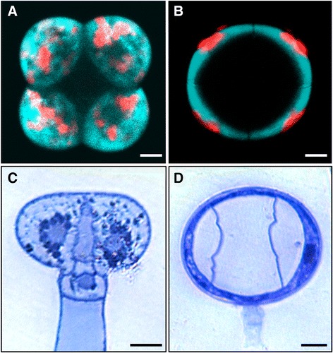
Fig. 2.

Fluorescence and bright field microscopy of type VI trichome heads from S. habrochaites and S. lycopersicum. a-b Fluorescence microscopy images of trichome heads of S. lycopersicum LA 4024 (a) and S. habrochaites LA 1777 (b). Bright field microscopy images of sections of type VI trichomes attached to the leaves and stained with toluidine blue from S. lycopersicum LA 4024 (c) and S. habrochaites LA 1777 (d). The horizontal bars correspond to 10 μm in all panels
