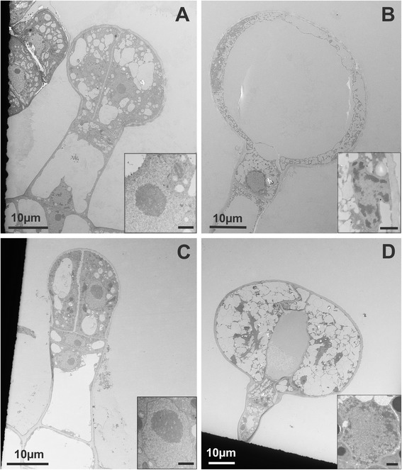
Fig. 7.

Electron microscopy of type VI trichomes at different stages of development. a-b S. habrochaites LA 1777 trichomes. a a young 4-cell stage trichome. b mature trichome. c-d S. lycopersicum LA 4024 trichomes. c: a young 2-glandular cell trichome. d Mature trichome. Inserts in a-d: ultrastructure of nuclei of glandular cells in the corresponding stage of development. The scale bar in the inserts represents 1 μM
