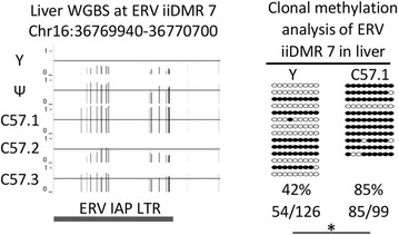
Fig. 4.

Methylation variability at ERV iiDMR 7 validated using an independent method. A screenshot of the WGBS methylation at ERV iiDMR 7 is shown for the five mice (left). On the Y-axis 0 represents no methylation, 1 represents 100 % methylation and the solid lines indicate the 50 % methylation position. The coordinates of the ERV iiDMR 7 overlaps an IAP LTR, indicated in dark grey. Methylation levels from clonal bisulphite sequencing (primer sequences are in Additional file 1: Table S5) on the two extreme samples (yellow and C57.1 mouse DNA) confirmed the differential methylation (right). Each sample is represented by at least 11 clones, filled in circles represent methylated CpGs from each sequenced clone. Asterisk indicates T test, p value <0.05
