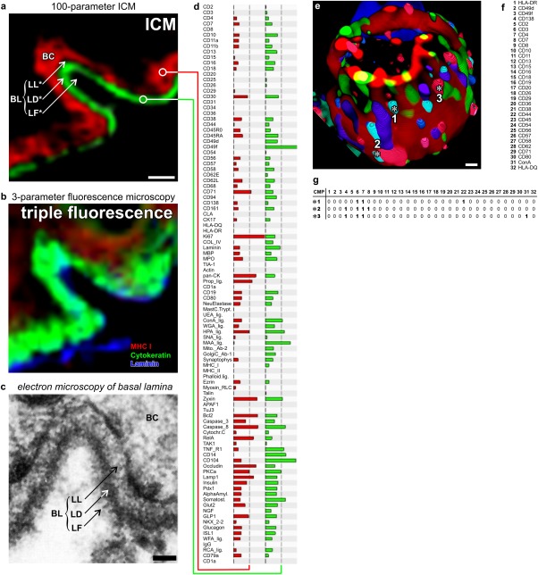
Figure 1.
Functional super resolution of large molecular networks. (a-d) Dermoepithelial junction in human tissue. Imaging cycler microscopy based discovery of molecular networks in situ 4. (a) Direct realtime protein profiling in 100-dimensional ICM data set using an algorithm based on the similarity mapping approach 19,20,22. Each data point has a PCMD of 256100. Note: sharp images at the junctional area discriminating between Lamina Fibroreticularis (LF), Lamina Densa (LD, green profile in d), Lamina Lucida (LL) and the basal ceratinocyte layer (BC, red profile in d), as known from transmission electron microscopy (c). (b) Same area as in (a), displaying traditional triple fluorescence imaging. (e) 3-dimensinal ICM imaging of distinct 32-component multi protein complexes on the cell surface of a blood T-lymphocyte 8. Multi protein complexes are composed of differential combination of 32 proteins/glycotopes listed in (f). (g) Examples are marked with asterisks (number 1 to 3) and detailed as CMPs with proteins present (1) or absent (0) together characterised as individual CMPs. Bars: 10 µm (a, b), 50 nm (c), 1 µm (e). A similar figure is featured in http://www.toposnomos.com/huto/tis.html. [Color figure can be viewed in the online issue, which is available at wileyonlinelibrary.com.]

