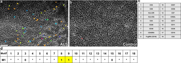
Figure 3.
ALS specific motif as predictive toponome biomarker visualized simultaneously in ALS PBMC (a) as directly compared with healthy control (b). The motif was revealed by fully automated ICM based co-mapping of 18 cell surface proteins (c) on isolated PBMC. The motif as a whole is composed of 200 distinct CMPs. The visual field of (a) shows cells, each of which displays one ALS-specific CMP out of the whole motif (different colours). The motif contains CD16 and CD45RA as lead proteins denoted (1 = lead protein, present in all CMPs) (d), while other proteins are either not associated (0 = anticolocated), or variably associated with the lead proteins (* = wild cards) (d, motif M1) 64–66. Courtesy of HUTO Project. Bar: 100 μm. [Color figure can be viewed in the online issue, which is available at wileyonlinelibrary.com.]

