
Figure 1.
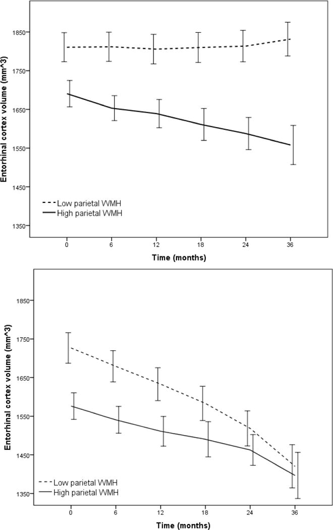
Rate of atrophy of entorhinal cortex volume stratified by total CSF t-tau level (Low t-tau top panel, High t-tau bottom panel) as a function of parietal white matter hyperintensity (WMH) volume dichotomized by mean log WMH value (for visualization purposes). Entorhinal cortex atrophy was particularly precipitous among individuals with low baseline CSF t-tau. Error bars are 95% confidence intervals.