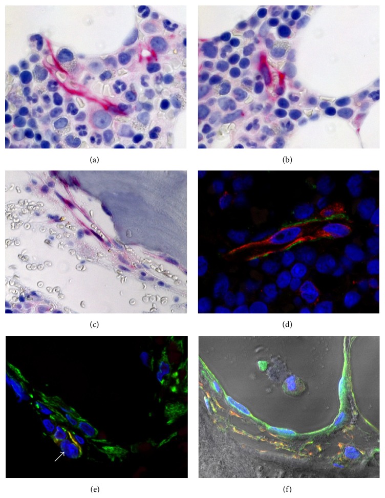
Figure 1.
Trephine BM biopsy obtained from a healthy control studied by immunohistochemistry (red) and immunofluorescence techniques (red, green, and coexpression signals: yellow) and visualized using brightfield (a–c) or laser scanning microscopy (LSM, (d–f)). (a, b, c) Nes+ cells (red) near arteriolar and sinusoidal blood vessels of the central BM areas (a, b) and (c) close the trabecular bone. (d) Localization of Nes+ (green) and CD146+ (red) cells around a small sinusoidal vessel in the central part of the BM. (e, f) Strong expression of CXCL12 (green) by endothelial and perisinusoidal cells that partially coexpress ((d), yellow) nestin (red) and synemin (green). Note perivascular CAR cell in (e) (arrow).

