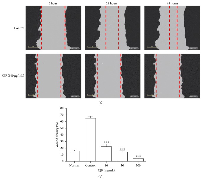
Figure 6.
(a) Confluent VSMCs were gently removed using the 96-pin WoundMaker to induce reproducible wounds and then the cells were treated with CJF for 1 h followed by PDGF-BB treatment. Images of wounded area were captured immediately (time 0) and 24 and 48 h after injury. (b) Finally, wound density was quantified as percentage of initial wound area that had been recovered with VSMCs. Results are shown as means ± SEM (n = 5). ∗ P < 0.05, ∗∗ P < 0.01, ∗∗∗ P < 0.001, significant difference versus control (PDGF-BB alone).

