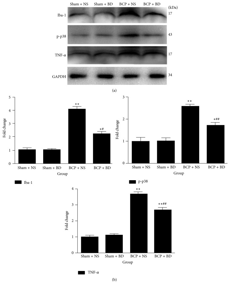
Figure 7.
Iba-1, p38 activation. and proinflammatory cytokine expression were reduced by intrathecal administration of BD1047. (a) Western blot analysis indicated that Iba-1, p-p38, and TNF-α expression were higher in the spinal cord of BCP rats on day 7. Intrathecal BD1047 significantly reduced the expression of these molecules in BCP rats compared with NS-treated BCP group. (b) Quantification of Iba-1, p-p38, and TNF-α expression level in the spinal cord (n = 4). Results are given as means ± SEM. ∗ P < 0.05, ∗∗ P < 0.01 versus sham + NS group; # P < 0.05, ## P < 0.01 versus BCP + NS group.

