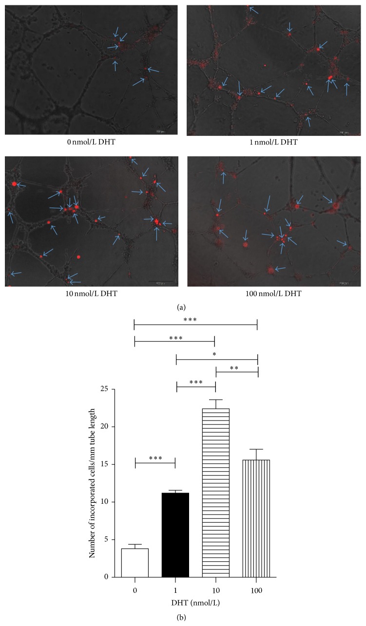
Figure 3.
In vitro incorporation assay by HUVECs and incorporated EPCs. DHT-treated or nontreated EPCs were tracked with DiI. DiI-labeled cells and HUVECs were seeded onto Matrigel-coated 96-well plates in 10% FBS/EBM2-MV. After 24 hours in culture, incorporation of each cell population into tube-like structures formed with HUVECs was evaluated under fluorescence microscopy. (a) Incorporated DiI positive cells were indicated by arrows. (b) Number of incorporated cells into tube-like structures was counted and averaged. All assays were triplicated and demonstrated similar results. Data are presented in mean ± SD format. ∗ P < 0.05 versus control group, ∗∗ P < 0.01 versus control group, and ∗∗∗ P < 0.001 versus control group.

