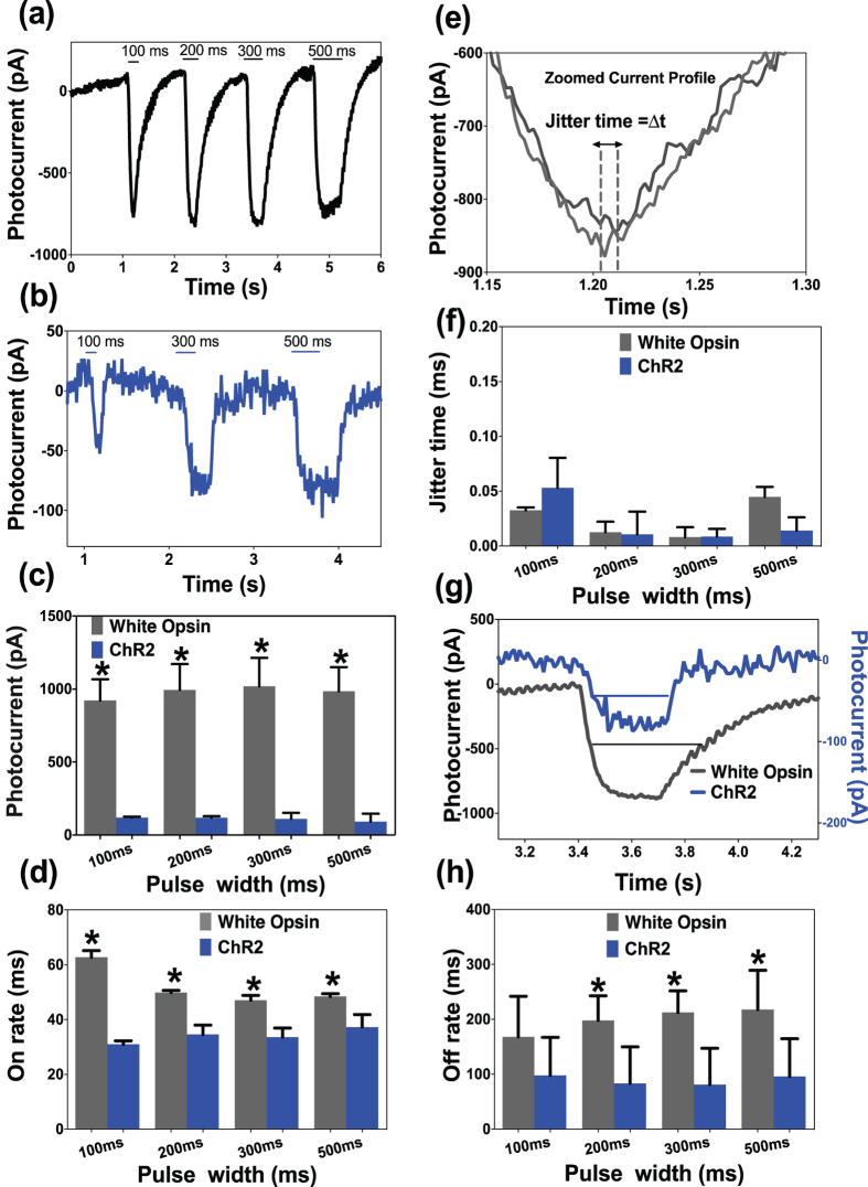
Figure 3. Dependence of peak current and on/off-rate on pulse-width of white-light stimulation of white-opsin expressing cells.
Representative inward current in white-opsin (a) and ChR2 (b) expressing HEK293 cells upon white-light illumination with different pulse widths at fixed intensity. (c) Measured inward peak-photocurrent in white-opsin or ChR2 expressing cells as a function of pulse width of white-light. N = 5/opsins, 15 sweeps. *p < 0.01 between white-opsin and ChR2. No statistically significant difference between different pulse widths. (d) Quantitative comparison of on-rate of white-opsin and ChR2 in response to white light at four different pulse widths at fixed intensity. N = 5/opsins, 15 sweeps. *p < 0.01 between white-opsin and ChR2. (e) Method for measuring jitter in inward photocurrent in same cell at fixed stimulation parameters. (f) Quantitative comparison of jitter time for white-opsin and ChR2 at different pulse width of white-light. (g) Zoomed photocurrent for white-opsin and ChR2 showing different off-rates. (h) Off-rate for white-opsin and ChR2 as a function of pulse width of white-light. N = 7/opsins, 21 sweeps *p < 0.05 between white-opsin and ChR2.

