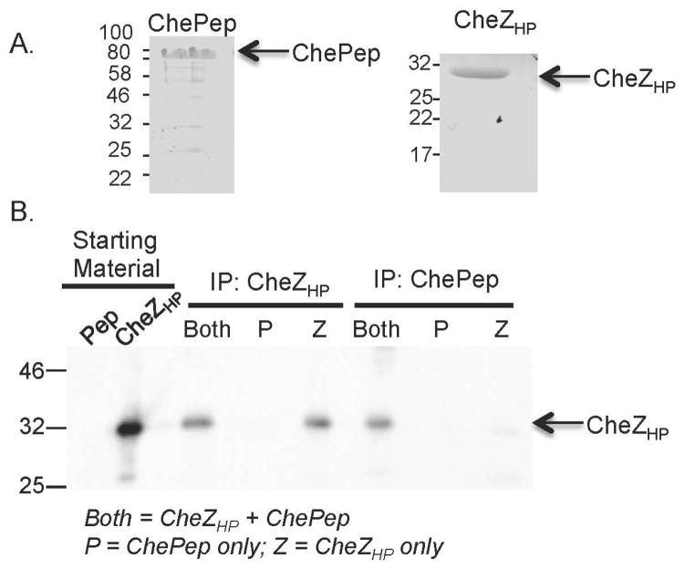
Figure 7.
CheZHP and ChePep interact directly. A. Coomassie-stained SDS-PAGE gel of purified ChePep (left) and CheZHP (right) proteins. Molecular weight in kilodaltons indicated at the left of each panel. B. Co-immunoprecipitation of CheZHP and ChePep, analyzed by western blotting of 10% SDS-PAGE gels with anti-CheZHP. From left to right: (1) Pep: the ChePep starting material (2) CheZHP: the CheZHP starting materials; (3–5) Immunoprecipitation (IP) with anti-CheZHP, incubated with a mixture of ChePep+CheZHP (both), ChePep (P) or CheZHP (Z); (6–8) IP with anti-ChePep, incubated with each set of proteins as in (3–5). The positions of ChePep and CheZHP are indicated on the right.

