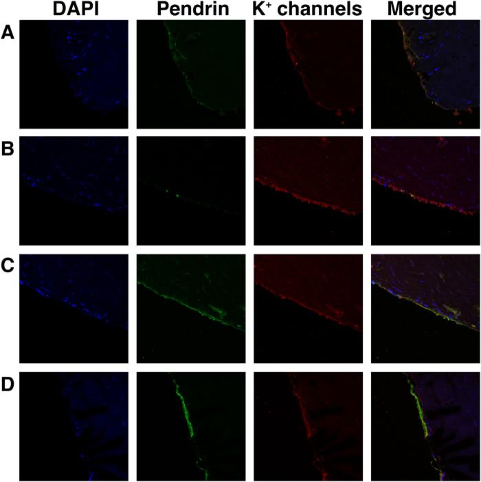
Figure 6. Immunofluorescent staining of K+ channels (KCNB1, KCNN2, KCNJ14, KCNK2, and KCNK6) identified by RT-PCR.
(a) KCNN2. (b) KCNJ14. (c) KCNK2. (d) KCNK6. The K+ channels except for KCNB1 were detected by immunofluorescent staining. Pendrin antibody was used for the validation of the ES epithelium because pendrin exists in mitochondria-rich cells of ES epithelial cells.

