
Figure 3. Characterization of the oligomeric state of the α6 subunit.
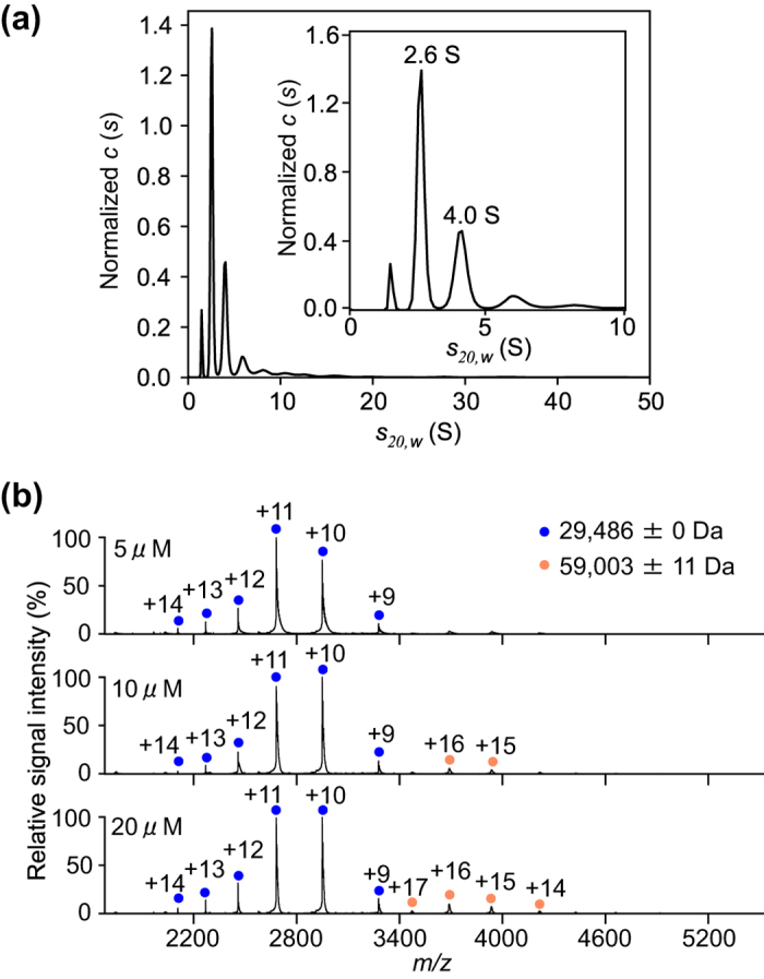
(a) Distribution of α6 sedimentation coefficients derived from SV-AUC experiments. Inset shows the enlarged view with s-ranging from 0–10 displaying the peaks corresponding to monomer (2.6 S) and dimer (4.0 S) of α6. (b) Mass spectra of α6 at 5, 10, and 20 μM under non-denaturing conditions. Blue and orange circles show the ion series of the α6 monomer and dimer, respectively.