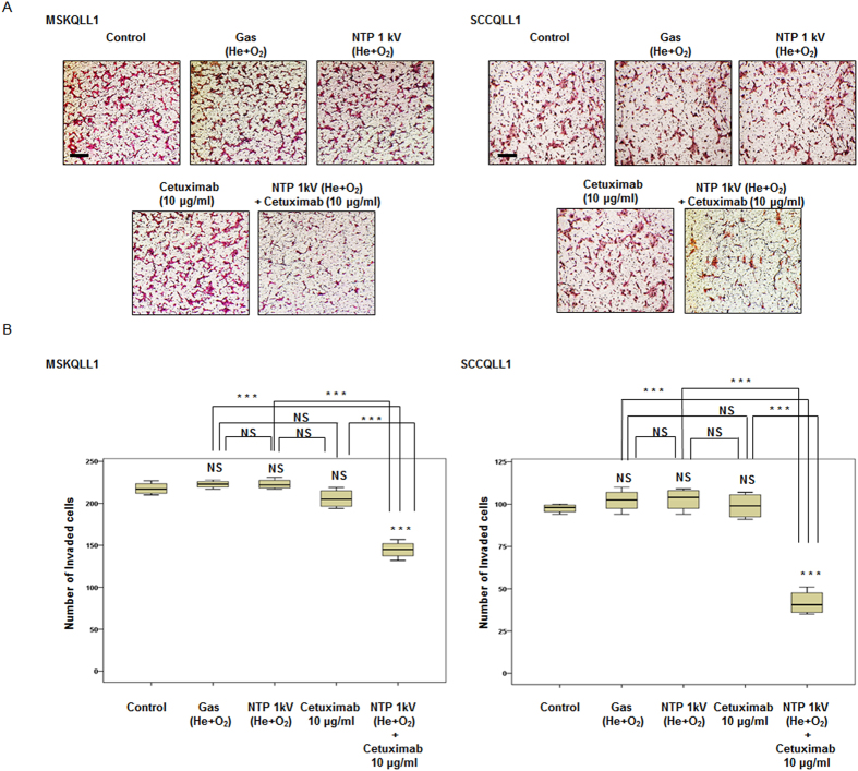
Figure 4. Combination of NTP with cetuximab decreased invasion by both MSKQLL1 and SCCQLL1 cells.
(A) Each cell line was seeded on a filter (pore size, 8 μm) coated with type I collagen in the upper chamber and exposed to gas (He plus O2 only), NTP (1 kV) and/or cetuximab (10 μg/ml). After 24 h, the cells attached to the lower section were stained with H&E. Each figure is representative of three experiments performed in triplicate. Scale bar = 50 μm. (B) To quantify invasion, stained cells in the lower chamber were counted using light microscopy (200×). The data represent the means ± S.D. of three independent experiments. NS, not significant; ***P < 0.001.

