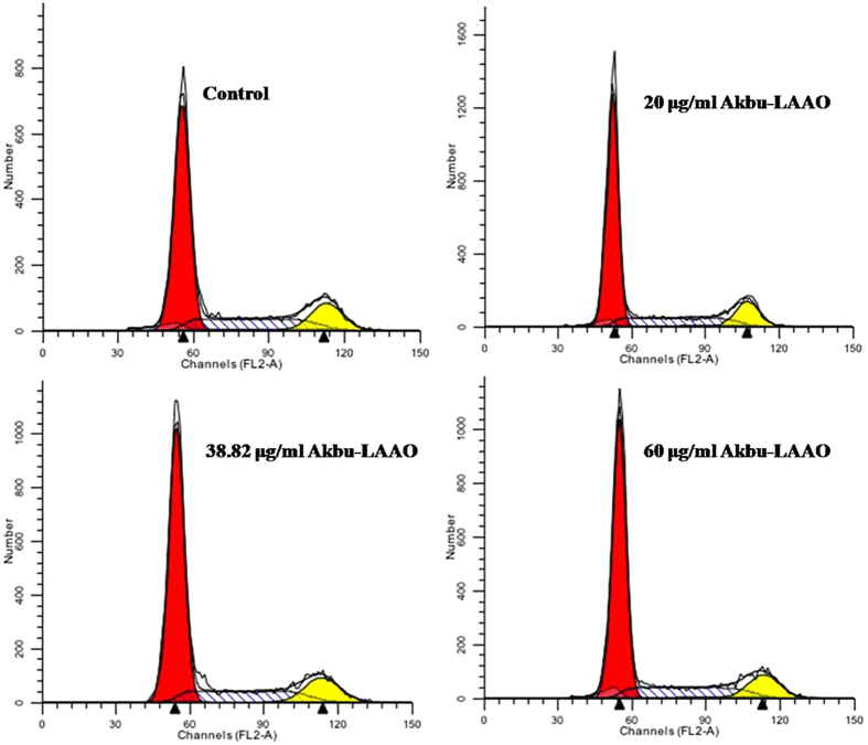
Figure 7. Akbu-LAAO treatment showed no effect on HepG2 cell cycle by flow cytometry assay.
Propidium iodide was used as the staining reagent. HepG2 cells were treated with 0, 20, 38.82 and 60 μg/mL of Akbu-LAAO for 24 h at 37 °C with 5% CO2. The cells stained with PI were subjected to flow cytometry foe measuring cell phase distributions.

