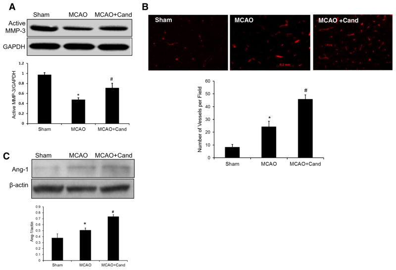
Fig. 4.
Low-dose candesartan treatment attenuates ischemia/reperfusion-induced neurovascular injury. a Representative and quantitative analysis of Western blots showing that expression of active MMP-3 was significantly decreased in the ipsilateral hemisphere 14 days after MCAO and salvaged by low-dose candesartan treatment. b Representative micrographs of laminin-stained brain sections collected 14 days after MCAO from saline- and candesartan-treated groups. Quantification of laminin-positive vessels shows higher vascular density in low-dose candesartan treatment as compared to saline-treated animals. c Representative and quantitative analysis of Western blots showing that low-dose candesartan treatment significantly increased Ang-1 expression after MCAO. Values are expressed as mean ± SEM [n=4–5/group, *P<0.05=MCAO + saline vs sham; MMP-3 (#P=0.0011), laminin (#P=0.0032), and Ang-1 (#P<0.0001)=MCAO + saline vs MCAO + cand]

